Sheridan
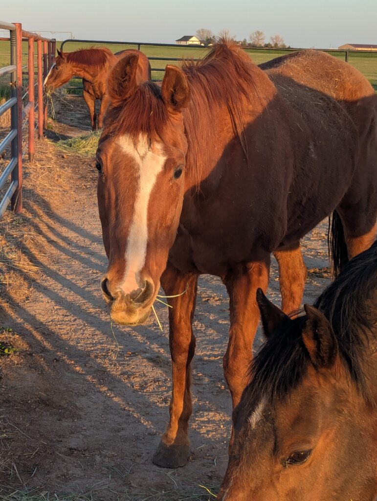
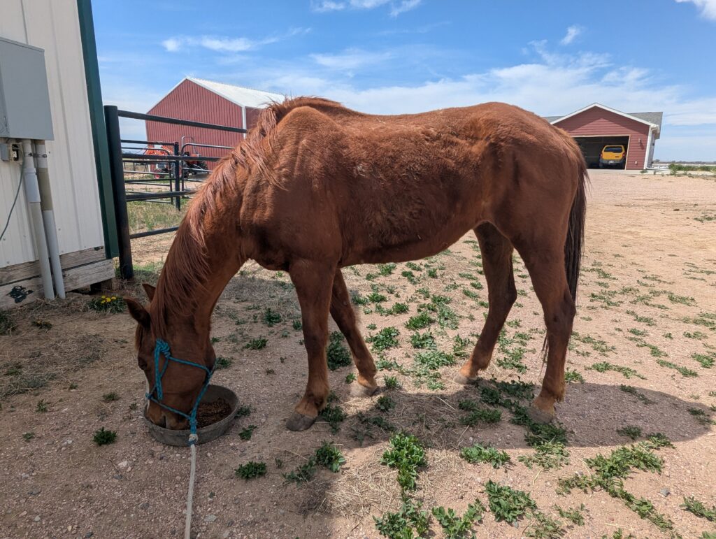
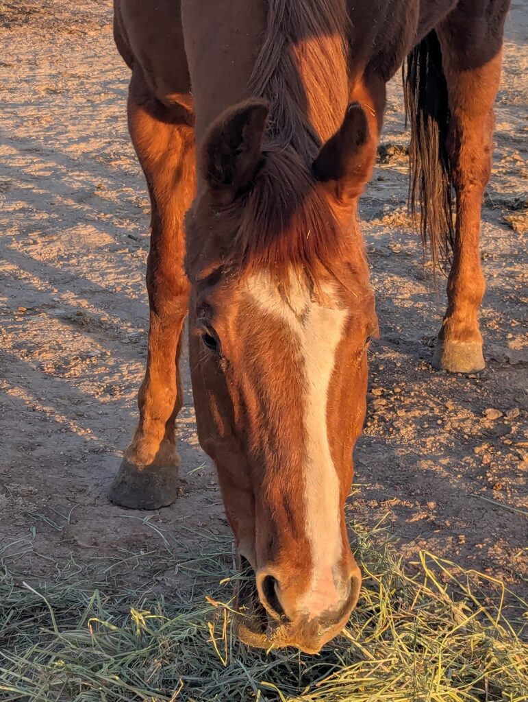
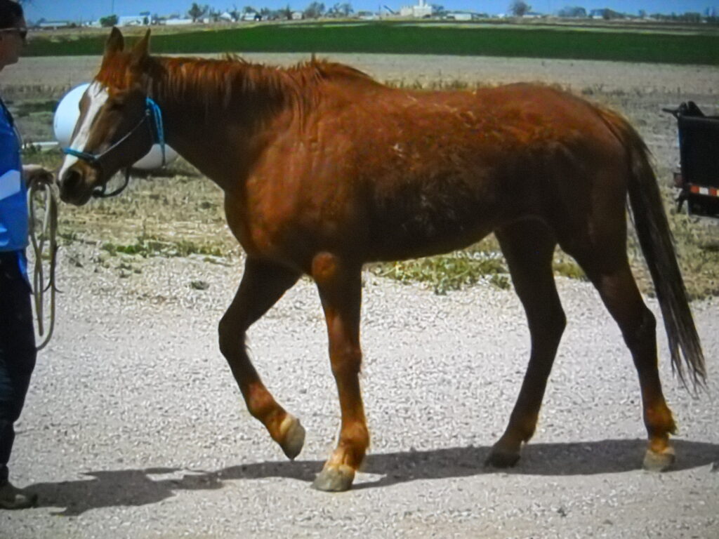
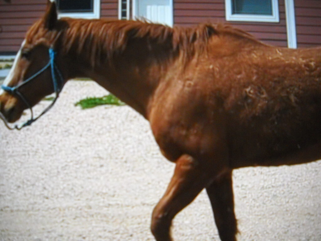
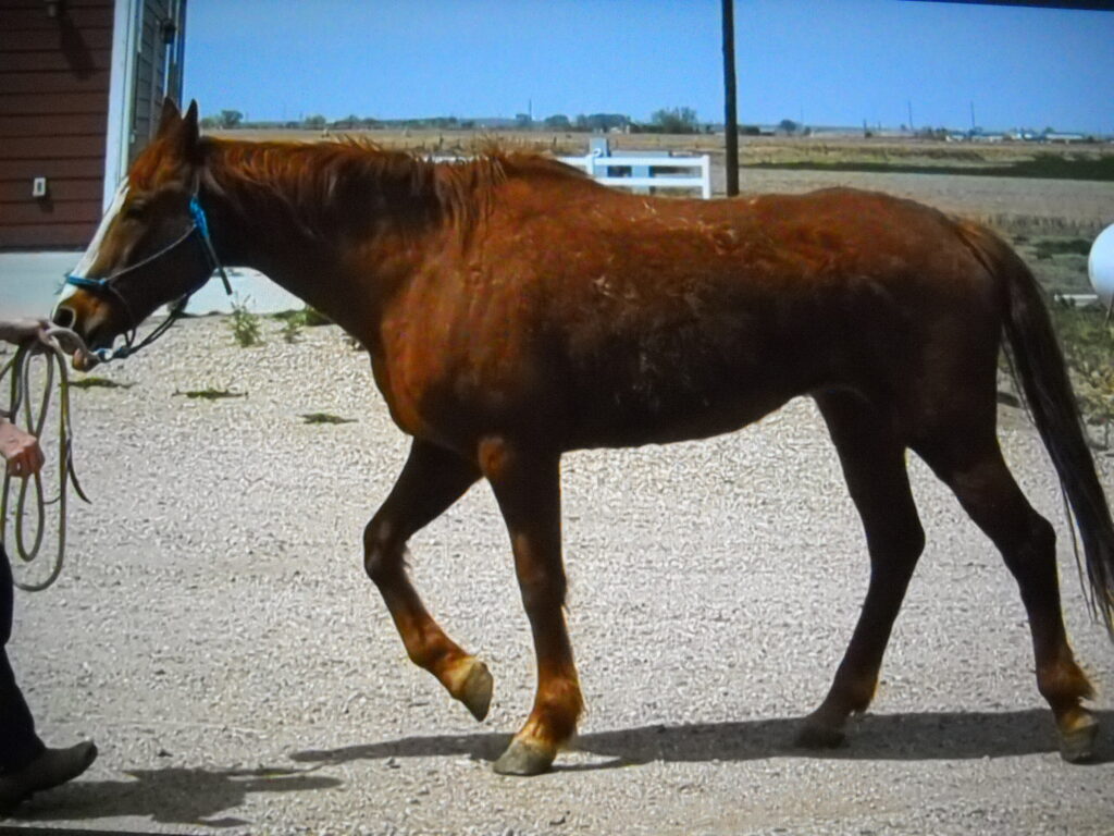
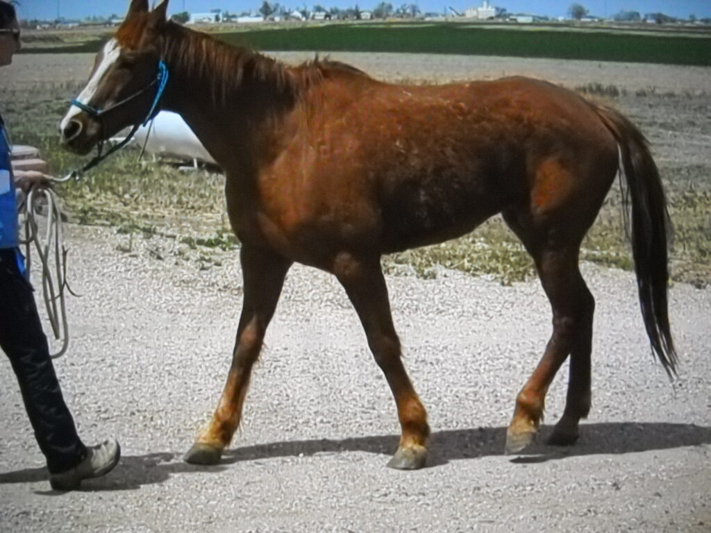
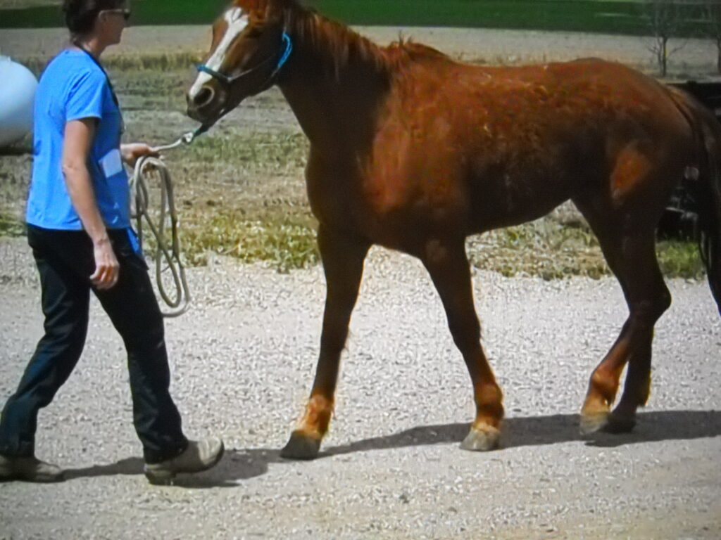
Sheridan was rescued to prevent her from entering the slaughter pipeline in April 2026. She’s a senior horse suffering from lameness which is due to older injury. She shows some neurological deficits and what looks like a possible tumor or similar growth (not a sarcoid) is under her right ear.
Sheridan also has atrophy present in her right hind, a very painful left hind hock and displays some stringhalt when moving.
Veterinary exam included lameness assessment along with Xray diagnostics. Some findings are below.
Sheridan walks with staccato lateral “stabbing” motion of both hind feet, and is grade 4/5 lame on the right hind leg. Both hind legs are positive from the Churchill pressure test, indicating a higher likelihood of hock pain.
A 2-view series dorsal and lateral views were taken of the right hock. While hock pain was suspected based upon exam findings of “stabbing” lateralized hind end gait and Churchill positive tests, flexions were not performed due to safety concerns, and so pain in these joints was not fully verified.
Moderate and chronic osteoarthritic (OA) changes are present in all four hock joints, both in the distal, low-motion joints (DIT, TMT) and high motion joints (tarsocrural and proximal intertarsal joints). The more proximal two joints, being high motion and therefore high use, can convey an amplified pain signal with small, to miniscule changes or clues on radiographs, while moderate to severe changes in the distal two joints may be manageable with conservative and medical interventions. The high motion joints have mild osteophyte changes seen on the dorsal tarsocrural joint on the dorsal aspect and sclerosis is present in the bone adjacent to articular cartilage of the talus.
Hock pain was likely a significant contributor to overall limb lameness score, with the possibility of other contributing factors elsewhere in the limb.
There is moderate valgus of the right hind pastern joint. Hind limb flexions were not attempted as part of this brief exam due to the degree of lameness and risk to veterinarian and/or an unknown horse and history.
An old scar crosses the forehead region and there is a firm lateralized dorsal swelling at the base of the right ear.
Post mortem radiographs were taken of the right oblique skull. The images suggested an enlarged tympanic bulla, which is a normal structure associated with the inner ear anatomy, but that might have been either congenitally enlarged or a traumatic/infectious/inflammatory event enlarged the bony “bubble” of bone. Unknown history with head scar and enlarged bulla, so these thoughts are merely conjecture. A 2-view series was also taken of the right hind hock.
After a full veterinary exam showed that her various health issues had no appropriate treatment for quality of life and her level of pain with further declining mobility would only worsen, she was humanely euthanized.
It is (yet again) another sad, inhumane and unnecessary situation where an innocent horse is used up, broken down, left to suffer and cast off to wring the last bit of money out of via a low-end auction.
More About Sheridan
- In Assessment
- Chestnut
- Mare
- 2003
- QH
- Colorado