Ollie
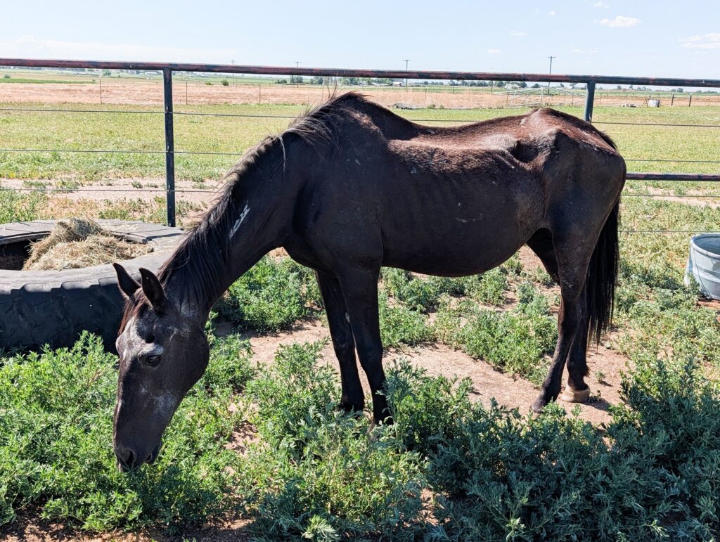
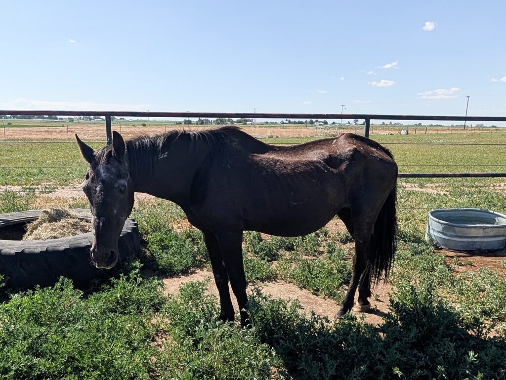
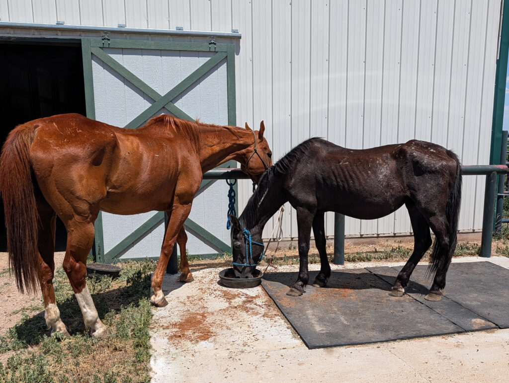
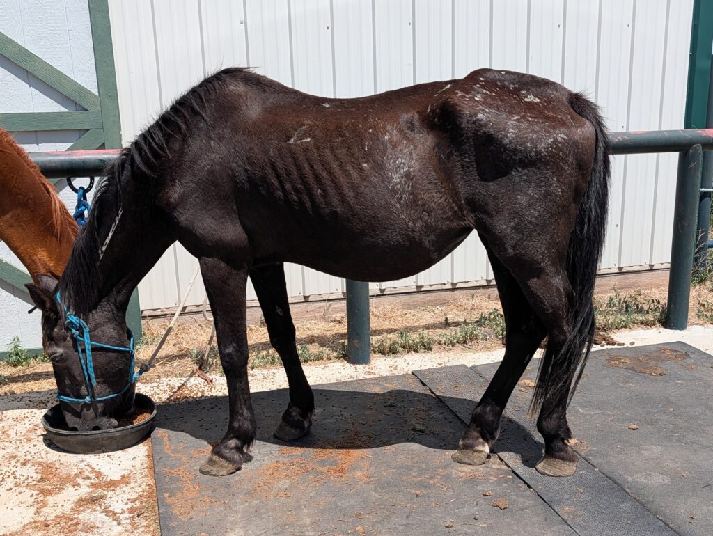
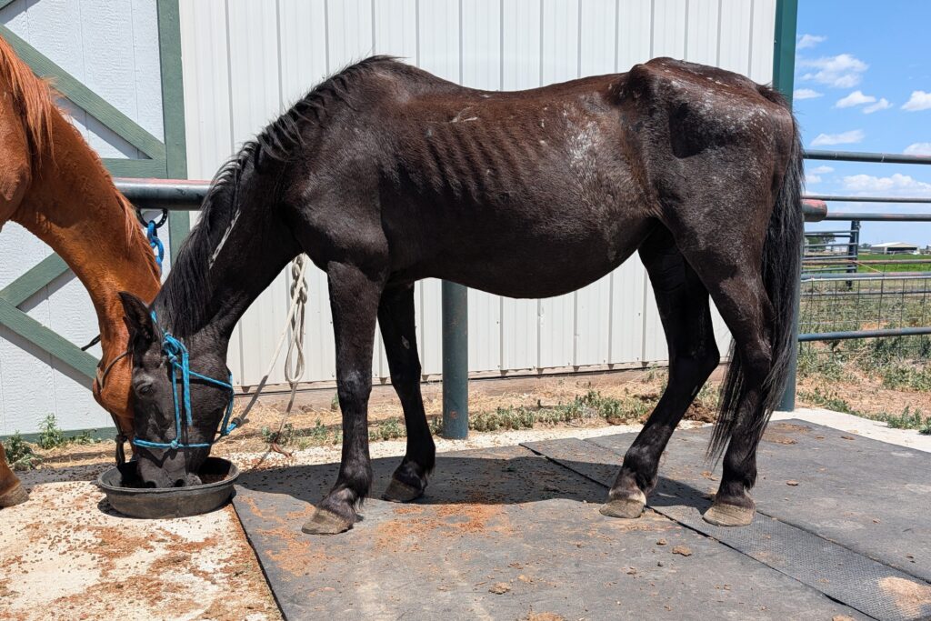
Ollie is an older, approximately 30+ year old emaciated mustang mare with a BLM freeze brand who was rescued during July 2026.
Her initial veterinary exam and evaluation provided both general and specific information:
“Ollie was emaciated,with a body condition score (BCS) of 2/9 with projecting skeletal structures such as individual dorsal spinal processes, tuber coxae, iliacus, and aged facial features. Both hind feet have misshapen hoof walls, with the right hind worse than left, with scooped-curvilinear dorsal hoof walls that have been bluntly cut off, bearing weight on her soles.
Ollie was rescued with a Thoroughbred companion (“Gulliver”) who showed likely signs of hepatotoxicity, likely induced by ingestion of toxic weeds on grass-insufficient pasture. If so, it’s highly suspect that Ollie would have ingested the same.
While Ollie did not have photosensitization lesions like Gulliver did, both of these genetically disparate horses did show similar signs of rapid emaciation and severe trauma to the dorsal thorax, lumbar, and sacral regions. It should be noted that Ollie’s companion (Gulliver) had white markings on his legs and a blaze on his face where photosensitivity reactions were easily seen. Ollie is black with no white markings.
Below is a brief summary of hepatotoxic plants:
a. Primary Photosensitization (Type I):Caused by the ingestion or contact of plants containing naturally photodynamic agents (e.g., St. John’s Wort, buckwheat, or spring parsley). The compounds circulate directly to the skin.
b. Hepatogenous Photosensitization (Type II/III): The most common and dangerous form in horses. It results from liver damage or disease. A healthy liver usually excretes phylloerythrin (a byproduct of chlorophyll digestion) via bile into the intestines. When the liver is damaged, it fails to clear this compound, which then accumulates in the skin and reacts violently to UV light.
Hepatotoxic Plants and Causes: Hepatogenous photosensitivity typically stems from liver failure triggered by the ingestion of toxic plants or mycotoxins. Common culprits include:Pyrrolizidine Alkaloid (PA) containing plants: Tansy ragwort, common groundsel, rattlebox, and houndstongue.
These cause chronic, progressive liver fibrosis like Clover: Known to cause severe hepatitis (liver inflammation) and secondary photosensitivity.
Blue-Green Algae (Cyanobacteria): Can cause acute, fatal liver necrosis. Symptoms typically present primarily on unpigmented (white) areas of the coat and hairless skin (muzzle, eyelids, and lower legs). Severe erythema (intense redness) and swelling, oozing and crusting lesions and ulceration as well as skin necrosis (tissue death) and sloughing off of skin.
Signs of underlying liver disease (e.g., jaundice/yellow mucous membranes) are restlessness, head shaking, and scratching due to intense pain and itching.”
Ollie is mildly lame on her right hind leg on firm gravel, with the right front lame at the trot only going on a right lunge circle. The right hind lameness remained the same in all directions and gaits.”
Due to Ollie’s advanced age, possible retirement future, unknown status of dentition but advanced age, and right limb lamenesses, a plan was made to provide further evaluation to obtain a better idea of lameness with radiography of feet, followed by an oral exam and possibly pulling blood to assess haptic function.
Xray images revealed the following:
“The right forefoot and left forefoot were each examined with lateral and DP views.
The right forefoot image shows osteoarthritis of the pastern and coffin joints, with a slight rotational change of the coffin bone of 1-2 degrees. The hoof wall shows a mild dished deflection in the lateral view about 4-6 months ago, indicating a possible laminitic episode.
The right hind foot shows marked changes which reflect the laminitic finding in the forefoot, but much more pronounced dishing of the dorsal hoof wall from an acute laminitic episode, widening of the white line, and rotation of the coffin bone of about 5 degrees. The distal tip of the coffin bone is severely deflected dorsally, indicating pedal osteitis. The tip is approximately 3-4 mm from the exterior hoof’s sole, a gravely insufficient thin sole, compounded focally by coffin bone rotation towards the ground.
Laminitis can be present in all four feet, especially in ponies or other insulin resistant or metabolic disease breeds (draft horses, gaited horses, mustangs). To find more prominent changes in a rear foot compared to a fore foot is highly unusual.
The hind limbs bear 30% of the body’s weight, so to see more dramatic rotation or sinking may indicate that either sinking had already occurred; the sole was thinner in this misshapen hind foot from the beginning, causing a chronically inflamed foot from pedal osteitis to be more prone to laminitis breakdown and separation from the hoof wall.”
Dental wise Ollie had missing teeth, sharp points and worn out molars, likely a combination of little to no dental care plus ageing.
It is not possible to fully know why Ollie’s body condition was so degraded, but in combination, lameness, inflammation, chronic conditions possibly inflamed by toxic weeds or dietary sugars as an effort to re-feed may have backfired such that naive past owner felt food meant pain, and slow starvation ensued. Also, absent teeth or rotting teeth in need of extraction can induce high levels of stress and consume vast amounts of calories, contributing to muscle and adipose wasting in short periods of time.
Just as with her bonded companion (Gulliver), there were no options to improve or manage Ollie’s most serious health conditions. She and Gulliver crossed the Rainbow Bridge together.
More About Ollie
- In Assessment
- Blue Roan
- Mare
- 1996
- American Mustang
- Colorado